
What Is an X-Ray?
An X-Ray is an imaging test that uses a small amount of radiation to create pictures of dense body structures. The images can show bones, joint spaces, and changes in spinal alignment. A qualified health professional reviews the images with your history and examination findings before making care decisions. For more detail, see First Chiropractic Appointment Checklist: What to Bring and Expect.
Quick Facts About X-Ray
Category
Diagnostic imaging
Used for
Viewing bones and joint spaces
Common confusion
It does not show all soft tissue problems
Also called
Radiograph, X-ray image
Often discussed with
Chiropractic Adjustment
Key Takeaways About X-Ray
- X-Rays show dense structures more clearly than soft tissue.
- Images can help guide decisions about chiropractic adjustment.
- An X-Ray does not show every cause of pain.
How X-Ray Works
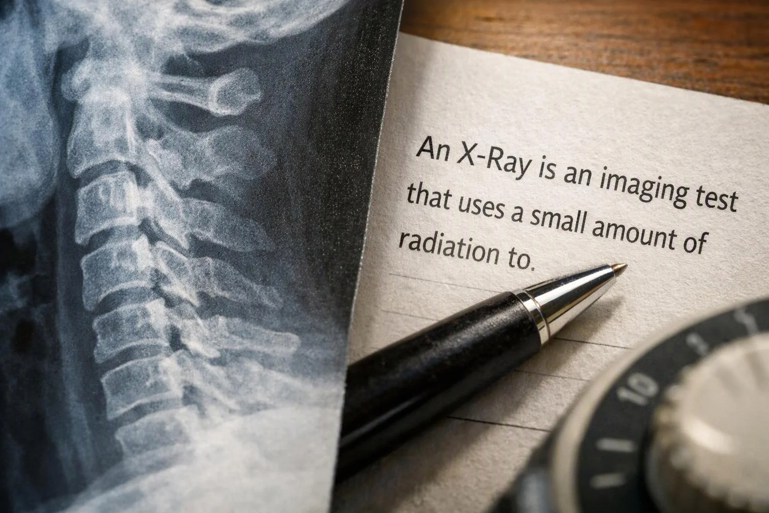
X-Ray imaging sends a controlled beam through part of the body. Dense tissue, such as bone, absorbs more of the beam. A detector records the pattern and creates an image. The image shows differences in bone shape, joint space, and alignment.
Related glossary terms: Cervical Spine, Lumbar Spine, Joint Mobility.
During a chiropractic evaluation, spinal alignment images may be reviewed with your examination findings. The views can help identify changes that affect how an adjustment should be planned. An X-Ray cannot show every muscle, ligament, or nerve problem, so the image is only one part of the clinical picture.
Chiropractic adjustment decisions also consider your symptoms, movement, and health history. A provider may review an existing image when it answers the clinical question, or request imaging through an appropriate medical channel when more information is needed.

From the Arrowhead Clinic - Lithia Springs team
When we assess a person for chiropractic adjustment, we review the examination findings before considering whether an X-Ray adds useful information. We look at the relevant area and connect the image with movement, symptoms, and health history. The image supports clinical judgment, but it does not replace the examination.
X-Ray in Practice: A Real-World Example
A person with back discomfort may have an X-Ray reviewed during an evaluation for chiropractic adjustment. The image may help the provider assess the lumbar spine and joint spacing alongside movement findings.
Related Services
Related Terms
Cervical Spine
Cervical Spine is the upper section of the spinal column that supports the head and protects…
Lumbar Spine
The lumbar spine is the lower section of the backbone between the rib area and the…
Joint Mobility
Joint Mobility is the amount and quality of movement available at a joint. It describes how…
Range Of Motion
Range Of Motion is the amount a joint can move in different directions. A shoulder's range…
Arrowhead Clinic - Lithia Springs
Have Questions About X-Ray?
Contact Arrowhead Clinic - Lithia Springs for practical guidance on X-Ray and related chiropractor work in Lithia Springs.